Enlarged Lungs Biography
(Source google.com)
In human anatomy, the bronchial
arteries supply the lungs with nutrition and oxygenated blood. Although there
is much variation, there are usually two bronchialarteries that run to the left
lung, and one to the right lung. The bronchial arteries supply blood to the
bronchi and connective tissue of the lungs. They travel with and branch with
the bronchi, ending about at the level of the respiratory bronchioles. They
anastomose with the branches of the pulmonary arteries, and together, they
supply the visceral pleura of the lung in the process. Note that much of the
blood supplied by the bronchial arteries is returned via the pulmonary veins
rather than the bronchial veins. Each bronchial artery also has a branch that
supplies the esophagus.
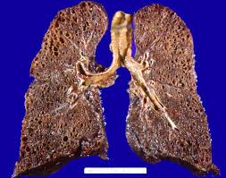
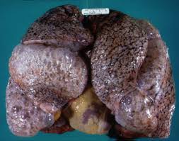
Sarcoidosis (from sarc meaning
"flesh", -oid, "like", and -osis, "diseased or
abnormal condition"), also called sarcoid, is a disease involving abnormal
collections of inflammatory cells (granulomas) that can form as nodules in
multiple organs. The granulomas are most often located in the lungs or its
associated lymph nodes, but any organ can be affected. Sarcoidosis seems to be
caused by an immune reaction to an infection or some other trigger (called an
antigen, which may be from one's environment) that continues even after the
initial infection or other antigen is cleared from the body. In most cases it
clears up by itself without any medical intervention, despite this some cases
do go on to affect the person long-term or become life-threatening and require
medical intervention, most often with medications. With an average mortality
rate of less than 5% in untreated cases. Treatment is usually designed to help
relieve the symptoms and hence do not directly alter the course of the disease.
This treatment usually consists of drugs
like ibuprofen oraspirin. In cases where the condition develops on a
progressive and/or life-threatening course the treatment is most often steroid
treatment with prednisone or prednisolone. Alternatively, drugs that are most
commonly used to treat cancer and suppress the immune system, such as
methotrexate, azathioprine and leflunomide may be used. In the United States it most commonly
affects people of Northern European (especially Scandinavian or Icelandish) or
African (especially African American) ancestry between the ages of 20 and 29,
although any race or age group can be affected. Japan
has a lower rate of sarcoidosis than the United States , although in these
people the disease is usually more aggressive in its course with the heart
often affected. Japanese individuals also have a different peak age for
sarcoidosis, namely 25–40 years of age.
It occurs more commonly in women too, with the female-to-male being
roughly 2:1, it also usually takes a more aggressive course in women.
In developing countries it often
goes misdiagnosed astuberculosis (TB) as its symptoms often resemble those of
TB. Sarcoidosis is a systemic inflammatory disease that can affect any organ,
although it can beasymptomatic and is discovered by accident in about 5% of
cases. Common symptoms, which tend to be vague, include fatigue (unrelieved by
sleep; occurs in 66% of cases), lack of energy, weight loss, joint aches and
pains (which occur in about 70% of cases), arthritis(14–38% of persons), dry
eyes, swelling of the knees, blurry vision, shortness of breath, a dry, hacking
cough, or skin lesions. Less commonly, people may cough up blood. The cutaneous
symptoms vary, and range from rashes and noduli (small bumps) toerythema
nodosum, granuloma annulare or lupus pernio. Sarcoidosis and cancer may mimic
one another, making the distinction difficult. The combination of erythema
nodosum, bilateral hilar lymphadenopathy, and joint pain is called Löfgren
syndrome which has a relatively good prognosis. This form of the disease occurs
significantly more commonly in Scandinavian patients, than in those of
non-Scandinavian origin.Localization to the lungs is by far the most common
manifestation of sarcoidosis. At least 90% of affected persons experience lung
involvement. Overall, about 50% develop permanent pulmonary abnormalities, and
5 to 15% have progressive fibrosis of the lungparenchyma. Sarcoidosis of the
lung is primarily an interstitial lung disease in which the inflammatory
process involves the alveoli, small bronchi and small blood vessels. In acute
and subacute cases, physical examination usually reveals dry rales. At least 5%
of persons will suffer pulmonary arterial hypertension. Less commonly, the
upper respiratory tract (including the larynx, pharynxand sinuses) may be
affected, which occurs in between 5 and 10% of cases. Sarcoidosis of the lungs
can be divided into four stages. Stage 0 — No intrathoracic involvement. Stage
I — Bilateral hilar adenopathy. Stage II — Pulmonary parenchyma involved. Stage
III — Pulmonary infiltrates with fibrosis.


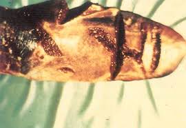
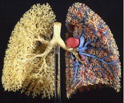
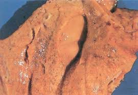
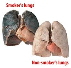


Enlarged Lungs Lungs Diagram of a Smoker after Smoking Cancer Anatomy And Heart Drawing Images AFter Smoking Wee of a Weed Smoker
Enlarged Lungs Lungs Diagram of a Smoker after Smoking Cancer Anatomy And Heart Drawing Images AFter Smoking Wee of a Weed Smoker
Enlarged Lungs Lungs Diagram of a Smoker after Smoking Cancer Anatomy And Heart Drawing Images AFter Smoking Wee of a Weed Smoker
Enlarged Lungs Lungs Diagram of a Smoker after Smoking Cancer Anatomy And Heart Drawing Images AFter Smoking Wee of a Weed Smoker
Enlarged Lungs Lungs Diagram of a Smoker after Smoking Cancer Anatomy And Heart Drawing Images AFter Smoking Wee of a Weed Smoker
No comments:
Post a Comment