Picture Of Lungs Biography
(Source google.com)
It has been long established that hostile personality traits are related to cigarette dependency and smoking cessation difficulties. Now UCI researchers have found that in people who have aggressive personalities nicotine triggers significant brain activity in the areas that help control social response, thinking and planning. In turn, non-hostile people showed no brain activity increases at all to nicotine. These findings suggest that some people are born with a predisposition to cigarette addiction and helps explain why quitting for some is practically impossible. It has been long established that hostile personality traits are related to cigarette dependency and smoking cessation difficulties. Now UCI researchers have found that in people who have aggressive personalities nicotine triggers significant brain activity in the areas that help control social response, thinking and planning. In turn, non-hostile people showed no brain activity increases at all to nicotine. These findings suggest that some people are born with a predisposition to cigarette addiction and helps explain why quitting for some is practically impossible. The ability of certain epithelial cells to use active-transport systems, as discussed above, enables them to absorb filtered material, such as glucose from the lumen of the intestine, which can then be circulated to the rest of the body. Cells are also able to endocytose other materials that are necessary for cell growth and signaling. For more information, see transcytosis. Some epithelial cells, such as the goblet cells, secrete fluids that are necessary for other processes such as digestion, protection, excretion of waste products, lubrication, reproduction, and the regulation of metabolic processes of the body. As part of its excretory role, certain epithelial cells secrete mucus, which lubricate the body cavities (i.e. peritoneum, pericardium, pleura, and tunica vaginalis) and passageways that they line. In the trachea, goblet epithelial cells secrete mucous which provides the lubrication to aid ciliated epithelial cells in sweeping bacteria and dust away from the lungs (Figure 6). In addition, type II alveolar cells excrete pulmonary surfactant, which decreases surface tension, allowing for normal lung function. Figure 6 shows an example of secretory cells in the fallopian tubes. Figure 6. This scanning electron microscope image depicts the ciliated epithelial cells that line the trachea. Some epithelial cells have cilia, which aid in moving substances in the lumen by creating a current via coordinated "sweeping" of the cilia (Figure 6). For instance, ciliated columnar epithelial cells are instrumental in the movement of the ovum through the Fallopian tubes to the uterus (Figure 7). Study: Take a group of people and, after standardized personality testing, divide them into two groups: a "high" hostile group characterized by more anger, aggression and anxiety as compared to a "low" hostile group. Include smokers and nonsmokers in both the "high" and the "low" hostile groups. Ask everyone to wear nicotine patches and then give them all brain scans. Obtain some interesting results. Nicotine triggered increased brain activity in the "high" hostile group -- whether they were smokers or not. (The "high" hostile smokers did need more nicotine to achieve a response comparable to the "high" hostile nonsmokers.) By contrast, there were no metabolic changes in the brain cells of the low-hostility participants. The results suggest that "high" hostile people respond to nicotine more than "low" hostile people.Conclusion: In people who have aggressive personalities, nicotine triggers significant brain activity in the areas that help control social response, thinking and planning. Comment: Does this mean that hostile people are more likely to start smoking in the first place and can be expected to have a harder time if they then decide to quit smoking. Study: Take a group of people and, after standardized personality testing, divide them into two groups: a "high" hostile group characterized by more anger, aggression and anxiety as compared to a "low" hostile group. Include smokers and nonsmokers in both the "high" and the "low" hostile groups. Ask everyone to wear nicotine patches and then give them all brain scans. Obtain some interesting results. Nicotine triggered increased brain activity in the "high" hostile group -- whether they were smokers or not. (The "high" hostile smokers did need more nicotine to achieve a response comparable to the "high" hostile nonsmokers.) By contrast, there were no metabolic changes in the brain cells of the low-hostility participants. The results suggest that "high" hostile people respond to nicotine more than "low" hostile people. Conclusion: In people who have aggressive personalities, nicotine triggers significant brain activity in the areas that help control social response, thinking and planning.
Comment: Does this mean that hostile people are more likely to start smoking in the first place and can be expected to have a harder time if they then decide to quit smoking? Pulmonary/Critical Care Medicine at Cedars-Sinai Medical Center and Professor of Medicine at UCLA. UCI study reveals why some people may be 'born to smoke' Nicotine study provides first results showing personality traits, brain activity and cigarette addiction link Why are some people hopelessly addicted to cigarettes, while others seemingly can quit at will? A UC Irvine College of Medicine study reveals for the first time the underlying brain mechanisms that link personality traits to nicotine addiction.

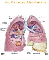

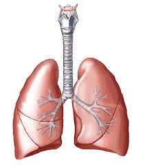

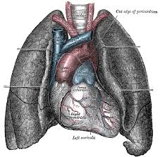

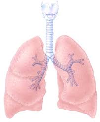


Picture Of Lungs Lungs Diagram of a Smoker after Smoking Cancer Anatomy And Heart Drawing Images AFter Smoking Wee of a Weed Smoker
Picture Of Lungs Lungs Diagram of a Smoker after Smoking Cancer Anatomy And Heart Drawing Images AFter Smoking Wee of a Weed Smoker
Picture Of Lungs Lungs Diagram of a Smoker after Smoking Cancer Anatomy And Heart Drawing Images AFter Smoking Wee of a Weed Smoker


