Lung Anatomy Biography
(Source google.com)
The human lungs are the organs of respiration in humans.
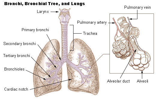
Humans have two lungs, a right lung and a left lung. The right lung consists of
three lobes while the left lung is slightly smaller consisting of only two
lobes (the left lung has a "cardiac notch" allowing space for the
heart within the chest). Together, the lungs contain approximately 2,400
kilometres (1,500 mi) of airways and 300 to 500 million alveoli, having a total
surface area of about 70 square metres (750 sq ft) to 100 square metres (1076.39
sq ft) (8,4 x 8,4 m) in adults — roughly the same area as one side of a tennis
court. Furthermore, if all of the capillaries that surround the alveoli were
unwound and laid end to end, they would extend for about 992 kilometres (616
mi). The lungs together weigh approximately 2.3 kilograms (5.1 lb), with the
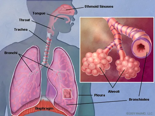
right lung weighing more than the left. The pleural cavity is the potential space between the two
serous membranes, (pleurae) of the lungs; the parietal pleura, lining the inner
wall of the thoracic cage, and the visceral pleura, lining the organs
themselves–the lungs. The respiratory system includes the conducting zone,
which consists of all parts of the airway that conducts air into the lungs. The parenchyma of the lung, only relates to the functional alveolar
tissue, but the term is often used to refer to all lung tissue, including the
respiratory bronchioles, alveolar ducts, terminal bronchioles, and all
connecting tissues. The lungs are located within the thoracic cavity, on either
side of the heart and close to the backbone. They are enclosed and protected by
the ribcage. The left lung has a lateral indentation which is shaped to
accommodate the position of the heart. The right lobe is a little shorter than
the left lung and this is to accommodate the positioning of the liver. Both
lungs have broad bases enabling them to rest on the diaphragm without causing
displacement. The left lung is divided into two lobes, an upper and a lower, by
the oblique fissure, which extends from the costal to the mediastinal surface
of the lung both above and below the hilum. There is also an area of the upper
lobe of the lung, specific to the left lung called the lingula, meaning little
tongue and is often referred to as the tongue. As seen on the surface, this
fissure begins on the mediastinal surface of the lung at the upper and
posterior part of the hilum, and runs backward and upward to the posterior
border, which it crosses at a point about 6 cm. below the apex. It then extends downward and forward over the costal surface,
and reaches the lower border a little behind its anterior extremity, and its
further course can be followed upward and backward across the mediastinal
surface as far as the lower part of the hilum.
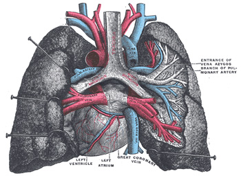
On the mediastinal surface, immediately above the hilum, is a well-marked curved furrow produced by the aortic arch, and running upward from this toward the apex is a groove accommodating the left subclavian artery; a slight impression in front of the latter and close to the margin of the lung lodges the left innominate vein. Behind the hilum and pulmonary ligament is a vertical furrow produced by the descending aorta, and in front of this, near the base of the lung, the lower part of the esophagus causes a shallow impression. The lungs are located within the thoracic cavity, on either side of the heart and close to the backbone. They are enclosed and protected by the ribcage. The left lung has a lateral indentation which is shaped to accommodate the position of the heart. The right lobe is a little shorter than the left lung and this is to accommodate the positioning of the liver. Both lungs have broad bases enabling them to rest on the diaphragm without causing displacement.
On the mediastinal surface, immediately above the hilum, is a well-marked curved furrow produced by the aortic arch, and running upward from this toward the apex is a groove accommodating the left subclavian artery; a slight impression in front of the latter and close to the margin of the lung lodges the left innominate vein. Behind the hilum and pulmonary ligament is a vertical furrow produced by the descending aorta, and in front of this, near the base of the lung, the lower part of the esophagus causes a shallow impression. The lungs are located within the thoracic cavity, on either side of the heart and close to the backbone. They are enclosed and protected by the ribcage. The left lung has a lateral indentation which is shaped to accommodate the position of the heart. The right lobe is a little shorter than the left lung and this is to accommodate the positioning of the liver. Both lungs have broad bases enabling them to rest on the diaphragm without causing displacement.
Lung Anatomy Lungs Diagram of a Smoker after Smoking Cancer Anatomy And Heart Drawing Images AFter Smoking Wee of a Weed Smoker
Lung Anatomy Lungs Diagram of a Smoker after Smoking Cancer Anatomy And Heart Drawing Images AFter Smoking Wee of a Weed Smoker
Lung Anatomy Lungs Diagram of a Smoker after Smoking Cancer Anatomy And Heart Drawing Images AFter Smoking Wee of a Weed Smoker

Lung Anatomy Lungs Diagram of a Smoker after Smoking Cancer Anatomy And Heart Drawing Images AFter Smoking Wee of a Weed Smoker

Lung Anatomy Lungs Diagram of a Smoker after Smoking Cancer Anatomy And Heart Drawing Images AFter Smoking Wee of a Weed Smoker

Lung Anatomy Lungs Diagram of a Smoker after Smoking Cancer Anatomy And Heart Drawing Images AFter Smoking Wee of a Weed Smoker

Lung Anatomy Lungs Diagram of a Smoker after Smoking Cancer Anatomy And Heart Drawing Images AFter Smoking Wee of a Weed Smoker

Lung Anatomy Lungs Diagram of a Smoker after Smoking Cancer Anatomy And Heart Drawing Images AFter Smoking Wee of a Weed Smoker
Lung Anatomy Lungs Diagram of a Smoker after Smoking Cancer Anatomy And Heart Drawing Images AFter Smoking Wee of a Weed Smoker

Lung Anatomy Lungs Diagram of a Smoker after Smoking Cancer Anatomy And Heart Drawing Images AFter Smoking Wee of a Weed Smoker

Lung Anatomy Lungs Diagram of a Smoker after Smoking Cancer Anatomy And Heart Drawing Images AFter Smoking Wee of a Weed Smoker
Lung Anatomy Lungs Diagram of a Smoker after Smoking Cancer Anatomy And Heart Drawing Images AFter Smoking Wee of a Weed Smoker
Lung Anatomy Lungs Diagram of a Smoker after Smoking Cancer Anatomy And Heart Drawing Images AFter Smoking Wee of a Weed Smoker
No comments:
Post a Comment