How Lungs Work Biography
(Source google.com)
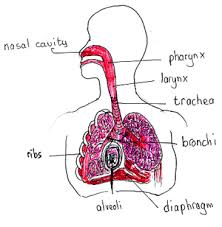
lungs are part of a group of organs and tissues that all work together to help you breathe. This system is called the respiratory system. The main job of the respiratory system is to move fresh air into and get waste gases out of the body. Oxygen, a basic gas, is needed by every cell in your body in order to live. The air that comes into the body through the lungs contains oxygen and other gases. In the lungs, the oxygen is moved into the bloodstream and carried through the body. At each cell in the body, the oxygen cells are exchanged for waste gas called carbon dioxide. The bloodstream then carries this waste gas back to the lungs where the waste gas is removed from the blood stream and then exhaled from the body. This vital process, called gas exchange, is performed automatically by the lungs and respiratory system.
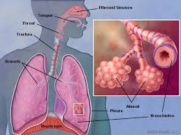
In addition to gas exchange, the respiratory system performs other roles important The SINUSES are hollow spaces in the bones of the head. Small openings connect them to the nose. The functions they serve include helping to regulate the temperature and humidity of air breathed in. The NOSE is the preferred entrance for outside air into the respiratory system. The hairs that line the wall are part of the air-cleaning system. Air also enters through the MOUTH, especially in people who have a mouth-breathing habit or whose nasal passages may be temporarily obstructed, as by a cold or during heavy exercise. The THROAT collects incoming air from the nose and mouth and passes it downward to the
The WINDPIPE (trachea) is the passage leading from the throat to the lungs. The windpipe divides into the two main BRONCHIAL TUBES, one for each lung, which subdivide into each lobe of the lungs. These, in turn, subdivide further. The right lung is divided into three LOBES, or sections. Each lobe is like a balloon filled with sponge-like tissue. Air moves in and out through one opening -- a branch of the bronchial tube. The left lung is divided into two LOBES. The PLEURA are the two membranes, actually one continuous one folded on itself, that surround each lobe of the lungs and separate the lungs from the chest wall. The bronchial tubes are lined with CILIA (like very small hairs) that have a wave-like motion. This motion carries MUCUS (sticky phlegm or liquid) upward and out into the throat, where it is either coughed up or swallowed. The mucus catches and holds much of the dust, germs, and other unwanted matter that has invaded the lungs. You get rid of this matter when you cough, sneeze, clear your throat or swallow. The smallest subdivisions of the bronchial tubes are called BRONCHIOLES, at the end of which are the air sacs or alveoli
The ALVEOLI are the very small air sacs that are the destination of air breathed in. The CAPILLARIES are blood vessels that are imbedded in the walls of the alveoli. Blood passes through the capillaries, brought to them by the PULMONARY ARTERY and taken away by the PULMONARY VEIN. While in the capillaries the blood gives off carbon dioxide through the capillary wall into the alveoli and takes up oxygen from the air in the alveoli.The DIAPHRAGM The THROAT collects incoming air from the nose and mouth and passes it downward to the
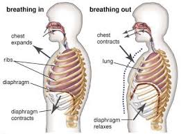
The WINDPIPE (trachea) is the passage leading from the throat to the lungs. The windpipe divides into the two main BRONCHIAL TUBES, one for each lung, which subdivide into each lobe of the lungs. These, in turn, subdivide further. The right lung is divided into three LOBES, or sections. Each lobe is like a balloon filled with sponge-like tissue. Air moves in and out through one opening -- a branch of the bronchial tube. The left lung is divided into two LOBES. The PLEURA are the two membranes, actually one continuous one folded on itself, that surround each lobe of the lungs and separate the lungs from the chest wall. The bronchial tubes are lined with CILIA (like very small hairs) that have a wave-like motion. This motion carries MUCUS (sticky phlegm or liquid) upward and out into the throat, where it is either coughed up or swallowed. The mucus catches and holds much of the dust, germs, and other unwanted matter that has invaded the lungs. is the strong wall of muscle that separates the chest cavity from the abdominal cavity. By moving downward, it creates suction in the chest to draw in air and expand the lungs. The RIBS are bones supporting and protecting the chest cavity. They move to a limited degree, helping the lungs to expand and contract.


How Lungs Work Lungs Diagram of a Smoker after Smoking Cancer Anatomy And Heart Drawing Images AFter Smoking Wee of a Weed Smoker
How Lungs Work Lungs Diagram of a Smoker after Smoking Cancer Anatomy And Heart Drawing Images AFter Smoking Wee of a Weed Smoker
How Lungs Work Lungs Diagram of a Smoker after Smoking Cancer Anatomy And Heart Drawing Images AFter Smoking Wee of a Weed Smoker
How Lungs Work Lungs Diagram of a Smoker after Smoking Cancer Anatomy And Heart Drawing Images AFter Smoking Wee of a Weed Smoker
How Lungs Work Lungs Diagram of a Smoker after Smoking Cancer Anatomy And Heart Drawing Images AFter Smoking Wee of a Weed Smoker
How Lungs Work Lungs Diagram of a Smoker after Smoking Cancer Anatomy And Heart Drawing Images AFter Smoking Wee of a Weed Smoker
How Lungs Work Lungs Diagram of a Smoker after Smoking Cancer Anatomy And Heart Drawing Images AFter Smoking Wee of a Weed Smoker
No comments:
Post a Comment